Figure 3.
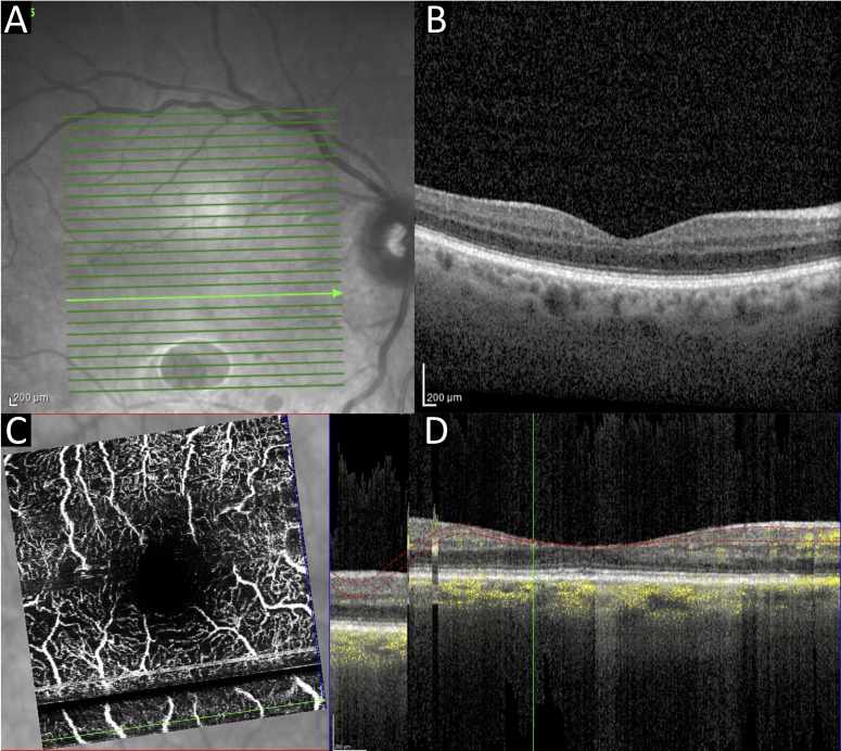
Case 2. Right eye OCT macula infrared (A) and cross-sectional view (B). OCTA macula en face view (C) and cross-sectional view (D). Poor ocular surface can be seen in the infrared image (A), and an alignment artefact arising from patient movement can be seen as a dark stripe on the OCTAs (C, D). OCT, optical coherence tomography; OCTA, OCT angiography.

