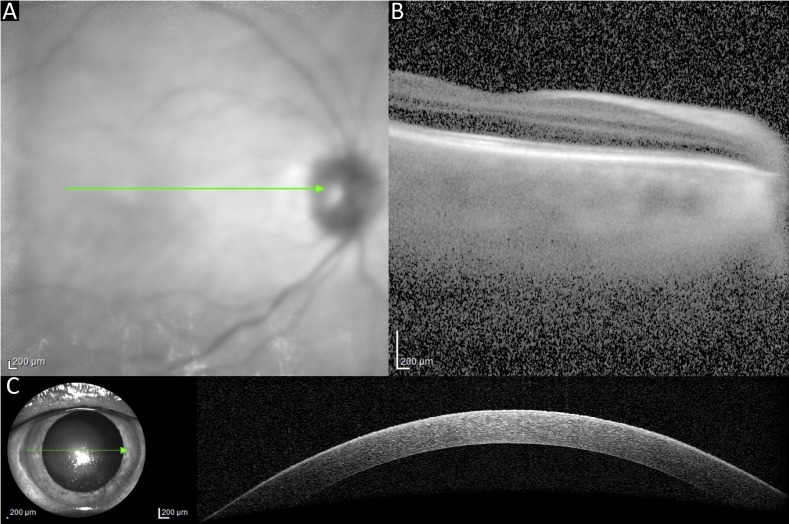
Figure 5.
Case 4. OCT macula (A, B) could only be acquired using a single line scan and the quality was poor. A single line macula was obtained, but the rest of the protocol was abandoned. A single line OCT scan of the anterior segment was obtained to assess integrity of the cornea (C). Green arrows shown in these images represent the area show in the corresponding cross-sectional views.

