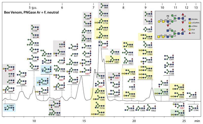
Figure 2. RP-HPLC of neutral N-glycans of honeybee venom.
N-glycans released by the combined use of PNGase F and Ar were subject to solid phase extraction, whereby the neutral-enriched fraction was eluted with 40% acetonitrile, prior to fluorescent labelling and chromatography on an RP-amide column; each fraction was collected and subject to MALDI-TOF MS. The annotations in the Symbolic Nomenclature for Glycans (see also key in grey box) are based on elution time, MS/MS and digestion data (see examples in Supplementary Figures 2 and 3). The column was calibrated in terms of glucose units. Phosphoethanolamine (PE)- or α-GalNAc-modified glycans in this pool are highlighted respectively in blue or green boxes, structures previously found on honeybee venom phospholipase A2 and hyaluronidase are in light grey boxes and those hybrid or biantennary forms reported by us in royal jelly in light yellow boxes; seven different isomers of Hex3HexNAc4Fuc2 are indicated by the m/z 1687 values in red. Due to their low abundance, neither the biantennary and Man4-5GlcNAc2-based hybrid glycans nor the PE-, β1,3-Gal and α1·4· alNAc-modified antennae were previously detected in honeybee venom.

