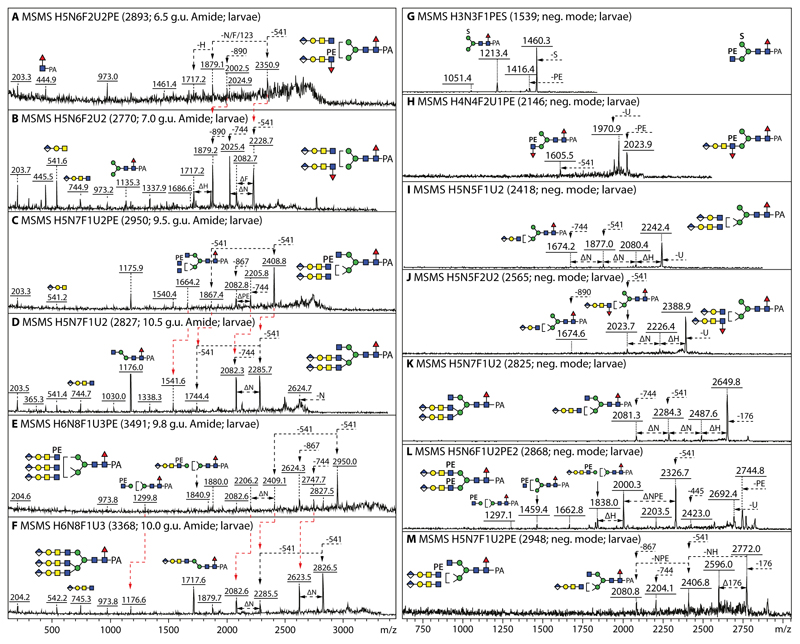
Figure 5. Positive and negative ion mode MALDI-TOF MS/MS of glucuronylated and zwitterionic N-glycans.
(A-F) Positive mode MALDI-TOF MS of larval bi- and tri-antennary glucuronylated glycans modified with or without phosphoethanolamine, whereby changes in fragmentation due to its absence or presence are indicated with red lines (Δm/z 123). (G-M) Negative mode MALDI-TOF MS/MS of glycans carrying either sulphate, phosphoethanolamine, glucuronic acid and/or fucose; in the negative mode, Y-ions resulting from serial losses are observed as long as they contain an anionic (GlcA or sulphate) or zwitterionic (PE) moiety. Panels D and K show the positive and negative mode MS/MS spectra of the same HexA2Hex5HexNAc7Fuc1 glycan (m/z 2827/2825). Losses of 541 (HexA1Hex1HexNAc1), 744 (HexA1Hex1HexNAc2), 890 (HexA1Hex1HexNAc2Fuc1) and 868 (HexA1Hex1HexNAc2PE1) from the multiple glucuronylated antennae are indicated; the annotations were aided by the pattern of losses (all panels) as well as the earlier RP-HPLC elution of the phosphoethanolamine-modified glycans (compare A and B, C and D, or E and F).

