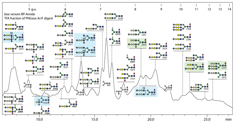
Figure 7. RP-HPLC of anionic N-glycans of honeybee venom.
PNGase F/Ar-released N-glycans were subject to solid phase extraction, whereby the anionic-enriched fraction was eluted with 40% acetonitrile/0.1% trifluoroacetic acid, prior to fluorescent labelling and chromatography on an RP-amide column; each fraction was collected and subject to MALDI-TOF MS. The annotations are based on elution time, MS/MS and digestion data (see Supplementary Figure 6); greyscale structures indicate the elution times of co-fractionating neutral glycans. The column was calibrated in terms of glucose units. Glycans with HexNAc3- or glucuronylated/phosphoethanolamine-modified antennae are highlighted respectively in green and blue boxes.

