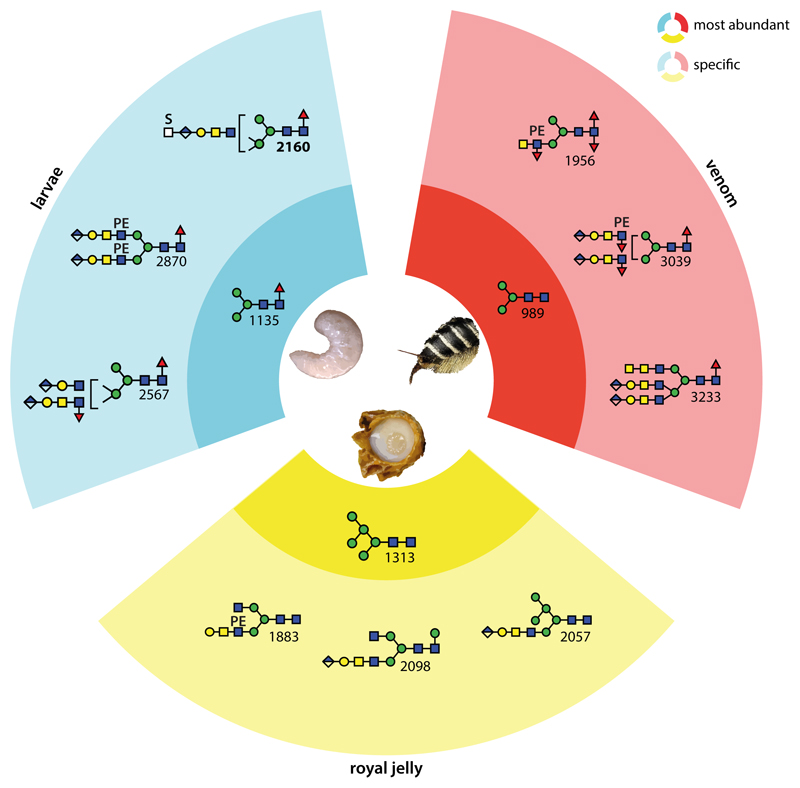
Figure 8. Summary of tissue-specific and overlapping N-glycan types in the honeybee.
The blue, yellow and red segments indicate structures specific to larvae, royal jelly and venom respectively; the darker sub-segments show the most abundant glycans in each sample, while the lighter sub-segments show three example sample-specific glycans. In the royal jelly, there is a dominance of hybrid structures and, as compared to larvae and venom, the glucuronylated antennae are ‘long’ (i.e., GlcAGalGalNAcGlcNAc rather than GlcAGalGlcNAc); processed venom glycans tend to be fucosylated on the core and antennae; in larvae, there is a bias towards β1,2- and β1,4-antennae, sometimes of different lengths. Interesting is that overall larvae and royal jelly share more structures with venom than royal jelly does with larvae. See also Supplementary Figures 9 & 10 and Supplementary Table 4 for an alternative summary of structures and for comparisons of MALDI-TOF MS profiles showing the major structures in each sample.

