
Fig. 5.
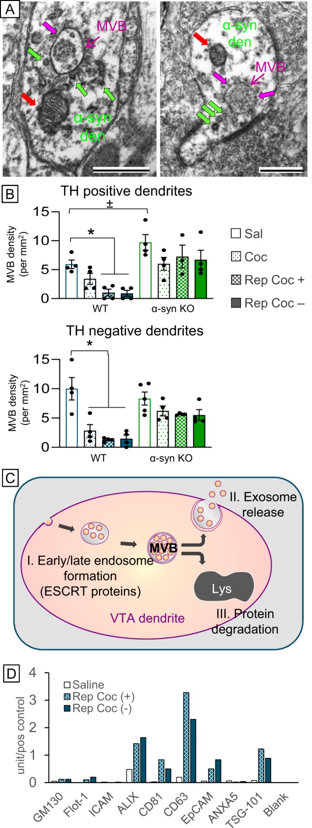
a Electron micrographs showing postsynaptic α-syn immunoperoxidase labeling on intracellular vesicles (green arrows) and around the outer membrane of MVBs (magenta arrows) and mitochondria (red arrows) in VTA dendrites. Scale bar = 500 nm. b Bar graphs showing that repeated cocaine administration decreased the presence of multivesicular bodies in dopamine (TH+) and non-dopamine (TH−) dendrites within the VTA, an effect that was diminished in α-syn KO mice (green bars). c Schematic diagram showing that decreases in MVBs can be caused by cocaine-mediated (I) dysfunction of MVB formation by ESCRT protein machinery, (II) increased exosome release, or (III) increased protein degradation by lysosomes. d Bar graph of antibody array quantification of isolated serum exosome samples from saline- and cocaine-treated mice showing increased ALIX, CD63, and TSG-101 immunoreactivity with repeated cocaine administration