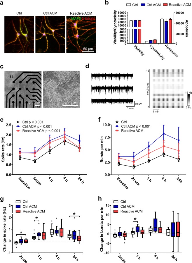
Figure 5.
Viability and functionality of neuronal cells after exposure to reactive astrocyte conditioned medium. (a) Immunocytochemical staining of hPSC-derived neuronal cells exposed to control medium (Ctrl), control ACM (Ctrl ACM) and reactive ACM. Neuronal cells stained positive for MAP2 (green) and β-tubulin (red). DAPI nuclear staining is shown in blue. (b) The reactive astrocyte conditioned medium did not show an effect on the viability, cytotoxicity or apoptosis of neuronal cells after 48 h of exposure (the data are representative of two experiments; in each experiment n = 3–6 per group). Statistical analysis was performed using independent-sample t-tests. (c) The effect of ACM on neuronal functionality was studied with microelectrode array (MEA) measurements. The phase-contrast images show neuronal cells cultured on MEAs. (d) Typical spontaneous activity in neuronal networks recorded from three electrodes over 5 minutes. The raster blot shows the typical network-wide activity from all 16 electrodes of one well over 5 minutes. (e) Spike rate (Hz) development for active electrodes before (baseline) and after different medium exposures (acute, 1 h, 4 h and 24 h). (f) Number of bursts per minute before and after different medium exposures. For the MEA data, n = 12 networks derived from one differentiation. The data are presented as the mean ± s.e.m., and statistical analysis was performed using the Friedman test. (g-h) To highlight significant differences between treatment groups, the data from panels e-f were reused and are presented as the changes in spike and burst rates compared to the baseline measurements. The data are shown as Tukey boxplots. Statistical significances was determined with the Mann-Whitney U test; * p < 0.05, ** p < 0.01, and *** p < 0.001.

