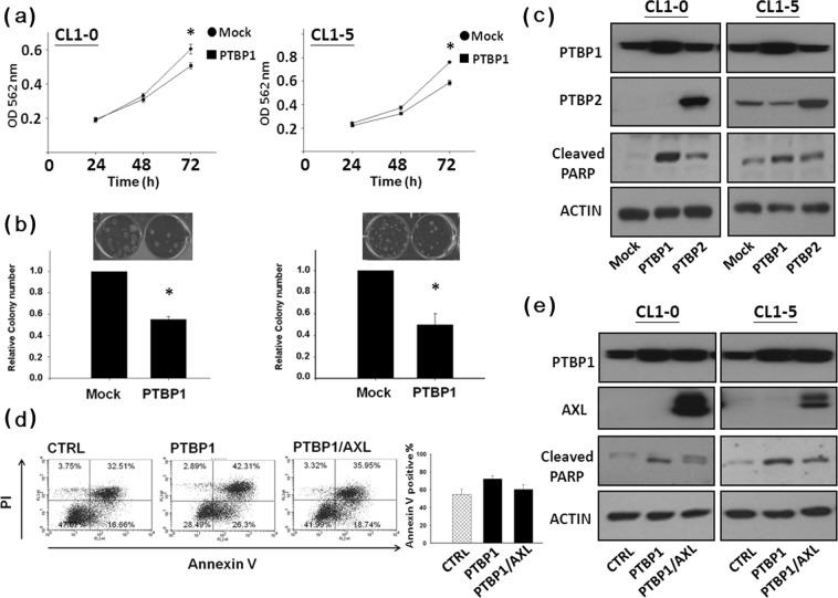
Figure 5.
AXL expression attenuates PTBP1-mediated apoptosis. Ectopic expression of PTBP1 in CL1-0 and CL1-5 cells inhibited cellular proliferation (A) and colony formation (B). (C) Cellular PARP is cleaved by PTBP1 over-expression. Cells were transfected with the mock, PTBP1 or PTBP2 vector, respectively. Western blot analysis of poly (ADP-ribose) polymerase (PARP) cleavage was performed 48 h later. Whole cell lysates were subjected to Western blotting using antibody against PTBP1, PTBP2 and PARP, respectively. Actin was used as an internal control for protein loading. (D) Over-expression of AXL counteracts the PTBP1-mediated apoptosis. After transfection, cells were allowed to express PTBP1 for 3 days prior to harvest. Apoptosis was determined by annexin V-FITC/PI double staining followed by flow cytometry analysis. The number in the lower right quadrant indicates the percentage of early apoptotic cells. The number in the upper right quadrant indicates the percentage of late apoptotic cells, and presented as mean ± SD. (E) Western blot analysis of poly (ADP-ribose) polymerase (PARP) cleavage. Whole cell lysates were subjected to Western blotting using antibody against PTBP1, AXL and PARP, respectively. Actin was used as an internal control for protein loading.

