Fig. 2.
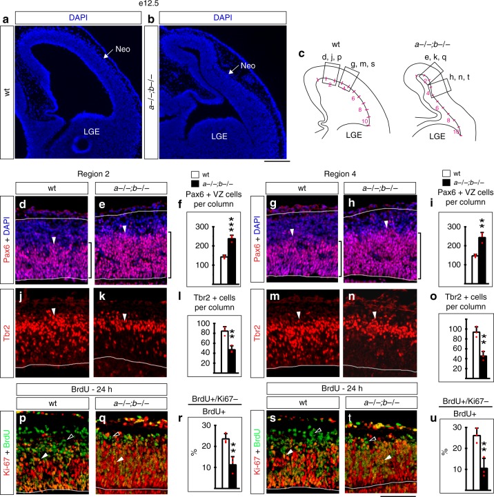
Accumulation of APs precedes gyrification of the Lmx1a−/−;b−/− cortex. Coronal sections at e12.5. a, b No evidence of cortical gyrification (outgrowths on outer cortical surface without bending of the corresponding ventricular surface) was detected in Lmx1a−/−;b−/− mutants at e12.5. Neo-neocortical primordium. LGE lateral ganglionic eminence. c Ventricular surface in wild-type and Lmx1a−/−;b−/− neocortical primordium (between the dorsal cortical bend and LGE) was subdivided into 10 equally sized regions (segments 1–10) and cells were quantified in segment 2 (d–f, j–l, p–r) and segment 4 (g–i, m–o, s–u). d–i Pax6+ APs (d, e, g, h, arrowheads) were increased in the number (f, i) (***p < 0.001 and **p < 0.01) and Pax6+VZ zone (bracket) was thicker in Lmx1a−/−b−/− mutants in both segments of the neocortical primordium. j–o Tbr2+ IPs (j, k, m, n, arrowheads) were reduced in the number (l, o) (**p < 0.01) in Lmx1a−/−;b−/− mutants in both segments of the neocortical primordium. p–u Mice were injected with BrdU 24 h prior to collecting embryos. p, q, s, t Arrowheads point to cells that re-entered the cell cycle (BrdU+/Ki67+ cells), open arrowheads—to cell that exited the cell cycle (BrdU + /Ki67− cells). A lower fraction of cells exited the cell cycle (the number of BrdU+/Ki67− cells divided by the number of BrdU+ cells) (**p < 0.01) in Lmx1a−/−;b−/− mutants in both segments of the neocortical primordium. All data are mean ± s.d., all p values are from two-tailed t-test, n = 3 embryos per genotype. Source data for panels f, i, l, o, r, u are provided as Source Data File. Scale bars: 200 μm (a, b); 100 μm (all other panels)

