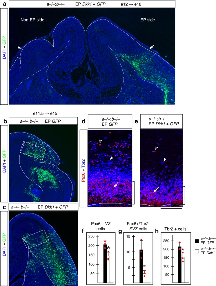
Fig. 4.
Electroporation of Dkk1 suppresses Lmx1a−/−;b−/− cortical gyrification. a An Lmx1a−/−;b−/− embryo in utero electroporated with Dkk1+ GFP at e12 and analyzed at e18. Early electroporation of Dkk1 into neocortical primordium restored lissencephaly (arrow), while the non-electroporated side remained gyrencephalic (arrowhead). b–h Lmx1a−/−;b−/− mutants electroporated with Dkk1+ GFP or GFP alone at e11.5 and analyzed at e15. Panels d, e are from sections adjacent to those shown in b, c and correspond to regions boxed in b, c. d–h In Dkk1-electroporated double mutants, the number of Pax6+ APs (arrows), Pax6+ /Tbr2− bRGs (open arrowheads) and Tbr2+ IPs (arrowheads) was reduced compared to GFP-electroporated double mutants (*p < 0.05, two-tailed t-test, n = 3 Dkk1-electroporated and n = 3 GFP-electroporated Lmx1a−/−;b−/− mutants). Bracket (d, e) indicates thickness of the Pax6+ VZ, which was also reduced in Dkk1-electroporated Lmx1a−/−;b−/−mutants. All data are mean ± s.d. Source data for panels f–h are provided as Source Data File. Scale bars: 400 μm (a); 200 μm (b, c); 100 μm (d, e)

