Fig. 6.
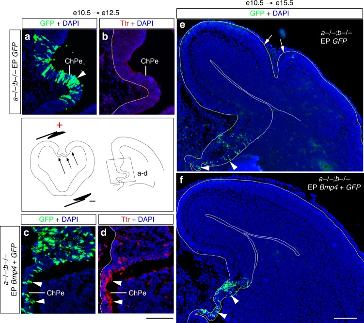
Bmp4 rescues DMe development and cortical gyrification in Lmx1a−/−;b−/− mutants. Bmp4 and/or GFP expressing plasmids were electroporated into DMe of Lmx1a−/−;b−/− embryos at e10.5 and analyzed at e12.5 (a–d) or e15.5 (e, f). To electroporate DMe, the positive electrode was placed against this region, as shown in the schematic below panel a. Negatively charged DNA moves toward the positive electrode (arrows) entering DMe. a–d Show DMe (a region boxed in the diagram below panel b). Immunohistochemistry revealed that Bmp4, but not GFP, induced Ttr in the Lmx1a−/−;b−/− ChPe (arrowheads in d). GFP+ ChPe cells (arrowheads in a, c) confirm successful electroporation of ChPe. e, f While GFP-electroporated Lmx1a−/−;b−/− mutants had cortical gyrification at e15.5 (e, arrows point to cortical gyri), electroporation of Bmp4 into DMe restored cortical lissencephaly (f). Arrowheads point to GFP+ cells in the DMe, showing specific electroporation of this domain. Scale bars: 100 μm (a–d); 200 μm (e, f)

