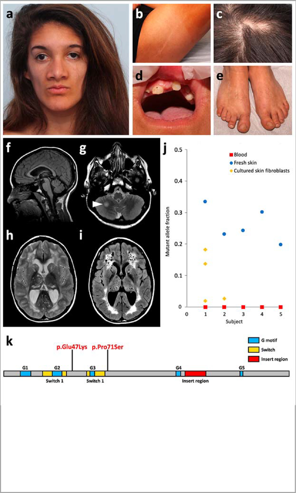
Figure 1 |. Main clinical features of RHOA-related mosaic ectodermal dysplasia and RHOA mutations.
a-e, Craniofacial appearance, linear hypopigmentation and other extracutaneous anomalies in subject S1. f-i, Brain MRI of subject S1 at 15 years. A second MRI, conducted 6 months later, did not show any significant change. Subjects S2 and S4 had similar but milder MRI abnormalities, including enlarged temporal horns of the lateral ventricles (Supplementary Fig. 2). Sagittal T1-weighted image revealed preserved midline structures (f). Axial T2-weighted images revealed a focal hyperintense lesion in the right hemisphere of the cerebellum (g, arrowhead), and diffuse cystic leukoencephalopathy with mildly enlarged lateral ventricles and cysts in the thalami and caudate nuclei (h). The leukoencephalopathy and presence of multiple cysts is confirmed on fluid-attenuated inversion recovery (FLAIR) sequences (i). j, Mutant allele fraction of RHOA mutations in the five subjects studied in WES or TUDS. k, Linear representation of RHOA and localization of the two mutations.

