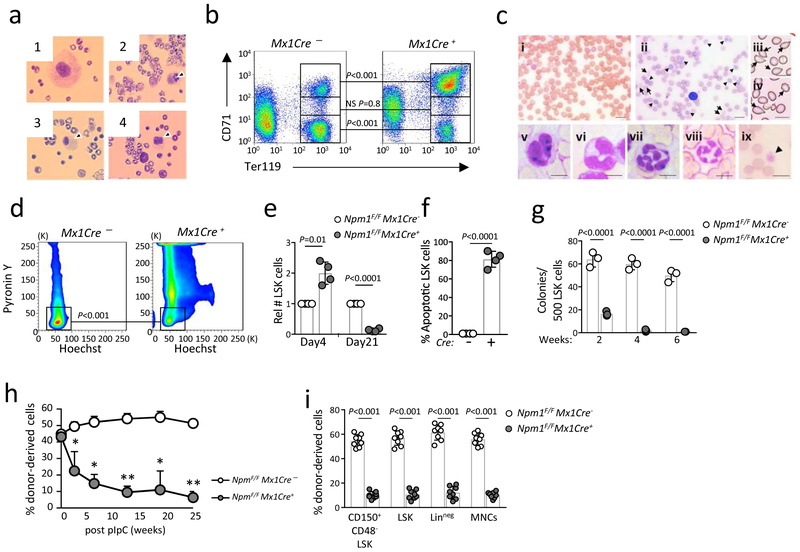
Figure 3. Acute deletion of Npm1 in adult mHSCs leads to BMF.
a-c, Acute loss of Npm1 in the hematopoietic system leads to human ribosomopathy phenotype. 7-10 days after pIpC injection, dysmegakaryopoiesis (a) and erythroid developmental defects (b) were observed, while 28 days after pIpC injection peripheral blood shows dysplastic features (c). a, Representative morphology (n = 20 biologically independent samples) of Npm1-deficient megakaryocytes (panel 2-4) 10 days after pIpC injection. b, Staining for TER119/CD71 positivity (n = 4 biologically independent samples) 10 days after Npm1 deletion is shown. c, Smears of peripheral blood of Npm1F/F;Mx1Cre− (c, panel i) or Npm1F/F;Mx1Cre+ mice (c, panels ii-ix) 4 weeks after pIpC administration show images of dysplastic erythroid cells (polychromasia, ii; poikilocytosis, iii,iv, arrowhead), dysplastic neutrophils (v,vi; hypersegmented neutrophils, vii,viii) and dysplastic platelets (giant platelet, ix). Scale bars, 10 μm. Shown are representative blood smear out of n = 20 independent biological samples. d, Npm1 deletion in adult HSCs leads to impairment of maintenance of quiescence. Percent of cells in G0 phase in Npm1-deleted HSCs (gated on LSK;CD48-;CD150+) 4 days after pIpC injection (n = 4 biologically independent samples). e, Relative number (± SD) of LSK cells 4 days and 21 days after Npm1 deletion, compared with pIpC-treated Npm1F/FMx1Cre− mice (n = 4 biologically independent samples per group). f, Npm1 deletion leads to increased apoptosis of LSK cells 21 days after pIpC injection. Data are presented as mean ± SD (n = 4 biologically independent samples per group). g, Results depict mean colony numbers ± SD/500 KSL cells (n = 3 biologically independent samples for each group). h-i, Reconstitution of donor cells in peripheral blood and bone marrow was monitored by staining blood cells with antibodies against CD45.2 (donor) and CD45.1. Data are presented as mean ± SD, (n = 8 biologically independent samples for each group h, and n = 8 biologically independent samples for each group, i). h, * P < 0.01, ** P < 0.0001. For all relevant panels, and unless otherwise stated, statistical significance was determined by one-tailed Student’s t test.

