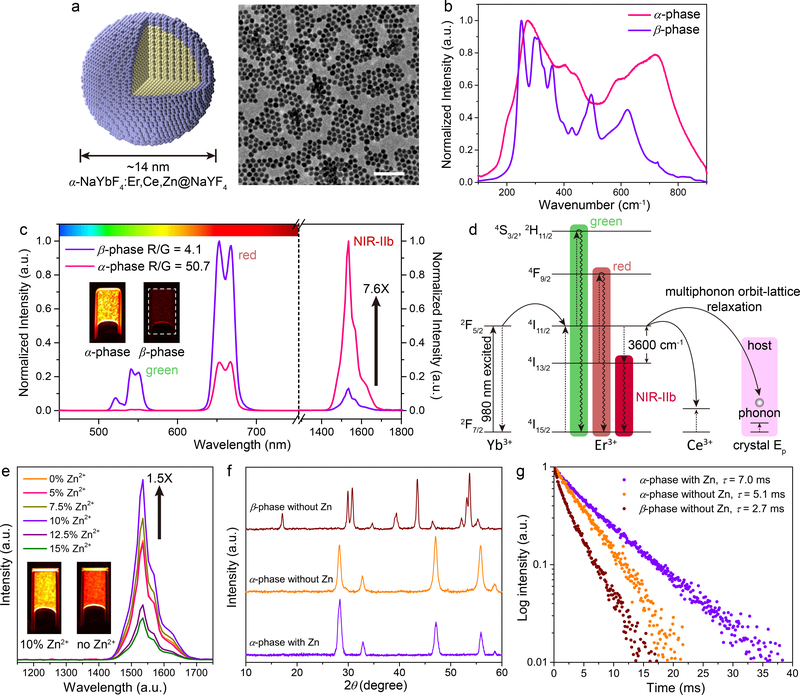
Figure 1: Ultra-bright ~ 1550 nm NIR-IIb luminescence of Zn doped α-ErNPs.
a, Schematic design of core-shell Zn doped α-ErNPs (left) and corresponding large scale TEM image (right, scale bar = 100 nm). b, Raman spectra of cubic-phase α-ErNPs and our previously reported hexagonal-phase ErNPs34. c, Upconversion and downconversion luminescence spectra of α-ErNPs and β-phase ErNPs. The insets showed NIR-IIb luminescence images of these two nanoparticles in cyclohexane. d, Simplified energy-level diagrams depicting the energy transfer involved in α-ErNPs upon 980 nm excitation. e, Downconversion luminescence spectra of Zn doped α-ErNPs with different Zn2+ concentration (0%, 5%, 7.5%, 10%, 12.5% and 15%, nominal doping concentration). The insets showed NIR-IIb luminescence images of α-ErNPs with 10% and 0% Zn2+ doping. f, g, XRD patterns f and lifetime decays g of cubic-phase α-ErNPs (10% Zn doping), cubic-phase α-ErNPs (0% Zn doping), and β-phase ErNPs. Similar results for n > 3 independent experiments.

