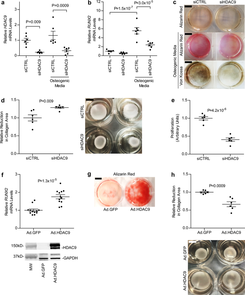
Figure 3 |. Vascular smooth muscle cell calcification, RUNX2 expression, proliferation, and contractility are affected by changes in HDAC9 expression.
a-c, Treatment of HASMCs (n = 6 biologically independent samples in each group) grown in osteogenic media with siHDAC9 (resulting in >75% knockdown of HDAC9 mRNA) (a) reduced RUNX2 mRNA levels by >50% (b) and prevented calcification, as evidenced by decreased Alizarin Red and von Kossa staining (c). In a and b, statistical comparisons were made using a two-tailed one-way ANOVA with Sidak’s test for multiple comparisons. In c, three independent experiments were performed with representative images shown. d, Reduced HDAC9 expression in HASMCs grown in collagen discs (right panel) resulted in a ~30% increase in contraction (left panel, n = 6 biologically independent samples in each group). e, Treatment of HASMCs with 20 nM siHDAC9 resulted in a 60% reduction in proliferation (n = 6 biologically independent samples in each group). f, Adenoviral expression of the 125-kDa isoform of HDAC9 fused to GFP in HASMCs was associated with a 75% increase in RUNX2 mRNA levels, when cells were harvested 8 days after viral transduction (n = 12 biologically independent samples in each group). Increased expression of HDAC9 was confirmed by Western blot (lower panel) using antibodies directed against HDAC9 and GAPDH (for a loading control). A full scan of the blot is in Supplementary Figure 4a. g, As shown by Alizarin Red staining, increased HDAC9 expression resulted in augmented calcification in HASMCs. Two independent experiments were performed with representative images shown. h, Increased HDAC9 expression also caused a 34% decrease (left panel, n = 6 biologically independent samples in each group) in contraction of HASMCs grown in collagen discs (right panel). In d-f and h, statistical comparisons were made using a two-tailed Student t test. Mean ± s.e.m. is depicted in all plots. For c and g, scale bar is 1 cm. For d and h, scale bar is 0.5 cm.

