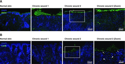
Figure 1. LecB localisation in chronically infected human wounds.
(A, B) Tissue sections of human infected wounds embedded in paraffin and stained for (A) P. aeruginosa (green) and for (B) LecB (green). Normal skin is used as negative control. Note: green signal in the upper left panel (A) is due to unspecific staining in the stratum corneum, not present in wounds. Rectangular squares refer to the zoomed area. Arrows point at LecB localised in the epidermal layers; arrowheads indicate LecB distributed in the dermis.

