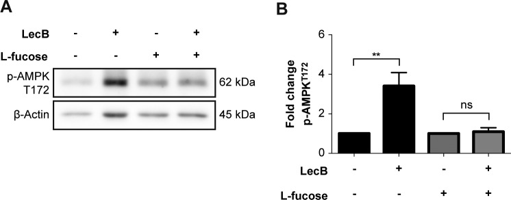
Figure S3. AMPK activation is blocked by L-fucose supplementation.
(A, B) Whole-cell lysates were analysed by Western blot to detect levels of pAMPK upon 4-h incubation with LecB (5 μg/ml) ± 30 mM L-fucose. The phosphorylated protein levels were normalised to actin. The graph reports the mean value ± SEM of N = 3 independent experiments. ** denotes P < 0.01, ns denotes not significant; one-way ANOVA was used for statistical analysis.

