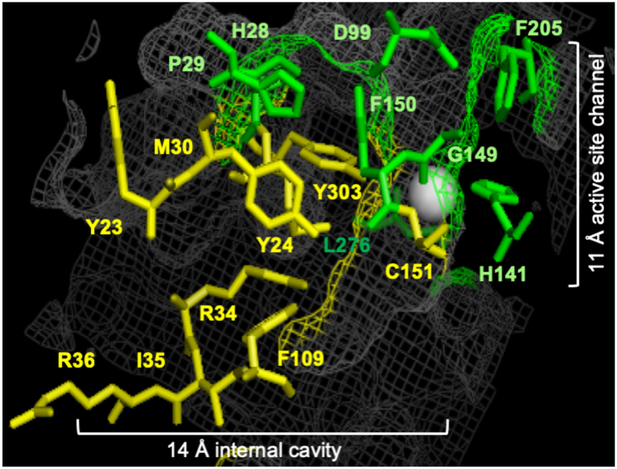
Figure 2.
HDAC1 trapping mutants. Crystal structure image of HDAC1 (PDB: 4BKX)[14b] where the seventeen amino acids in the 11 Å active site channel (green) and 14 Å internal cavity (yellow) of HDAC1 that were alanine mutated are shown as ball and stick structures. The HDAC1 structure is shown as grey mesh. The metal ion required for catalysis is shown as a white sphere. Image created with the Pymol molecular graphics system (version 1.3, Schrödinger, LLC).

