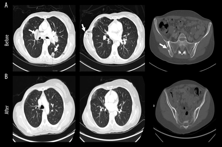
Figure 2.
Dramatic radiological response after chemo-immunotherapy. Computed tomography chest, abdomen and pelvis showing: (A) pretreatment; an increase in size of metastatic lymph nodes in the mediastinum and hila, multiple pulmonary metastatic lesions (left and middle) and a newly developed lytic lesion with soft tissue component in the right iliac bone (right), (B) post treatment; an excellent response with no suspicious pulmonary nodules (left and middle) and disappearance of right iliac bone soft tissue mass (right). Lesions are indicated in white arrows.

