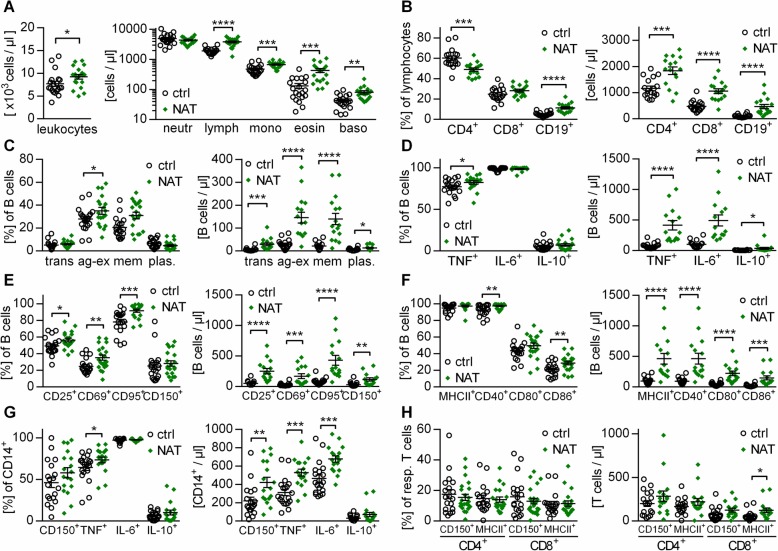
Fig. 2.
Natalizumab treatment increases the expression of activation markers and TNF production in B cells. Peripheral blood mononuclear cells (PBMC) were isolated from controls (n = 19; circles) or natalizumab (NAT; n = 20; diamonds)-treated multiple sclerosis patients. Cells were stained with the respective antibodies and analyzed using flow cytometry. Each dot represents the value of an individual patient; bars indicate mean ± standard error of the mean; *P < 0.05; **P < 0.01; ***P < 0.001; ****P < 0.0001; unpaired t test for cross-sectional data. a Total leukocyte counts and neutrophil, lymphocyte, monocyte, eosinophil, and basophil counts were determined from whole blood. b, c Cell frequencies and absolute cell counts were determined for b CD4+ and CD8+ T cells and CD19+ B cells as well as for c B cell subsets such as transitional B cells (trans), antigen-experienced B cells (ag-ex), memory B cells (mem), and plasmablasts (plas.). d PBMC were stimulated with 1 μg/ml CpG for 12 h, followed by 4 h of 500 ng/ml ionomycin / 20 ng/ml phorbol 12-myristate 13-acetate (PMA) stimulation. We determined the frequency as well as the absolute number of B cells producing tumor necrosis factor alpha (TNF), interleukin-(IL-)6, and IL-10. e, f Frequency and absolute number of B cells expressing markers for activation (e) and antigen presentation (f) were determined after 20 h of 2 μg/ml CpG stimulation. g PBMC were pre-stimulated for 12 h with 1 μg/ml CpG, followed by 4 h of 500 ng/ml ionomycin / 20 ng/ml PMA stimulation. We determined the frequency and absolute number of myeloid cells (CD14+) producing TNF, IL-6, or IL-10. CD150 expression was determined after 20 h of incubation with 100 pg/ml lipopolysaccharides (LPS). h PBMC were incubated with 100 pg/ml LPS for 20 h and determined their expression of CD150 and major histocompatibility complex class II (MHC-II)

