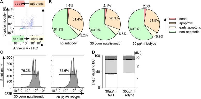
Fig. 5.
Neither apoptosis nor proliferation of B cells is affected by in vitro natalizumab exposure. Peripheral blood mononuclear cells (PBMC) were collected from healthy donors (n = 6). a, b PBMC were incubated in vitro for 72 h with no antibody, 30 μg/ml natalizumab (NAT), or an IgG4 isotype control antibody. To evaluate apoptosis of CD19+ B cells, cells were stained with anti-CD19 antibody, propidium iodide (PI), and annexin V (AV). Non-apoptotic B cells (PI− AV−), early apoptotic B cells (PI− AV+), apoptotic B cells (PI+ AV+), and dead B cells (PI+ AV−) were distinguished. a Exemplary dot plot and b respective pie charts showing the mean frequency of the analyzed parameters. c, d B cells were isolated from PBMC using magnetic-activated cell sorting (MACS). After carboxyfluorescein succinimidyl ester (CFSE) staining, they were stimulated with anti-human IgG and IgM F(ab)2 fragments (20 μg/ml), anti-human CD40 (10 μg/ml), CpG (0.5 μg/ml), and interleukin (IL)-21 (50 ng/ml) for 72 h in the presence of 30 μg/ml of natalizumab or IgG4 isotype control antibody. Frequency of dividing cells was determined by CFSE dilution using flow cytometry. c Representative histograms (d) and frequency of B cells showing 1, 2, or > 2 divisions (div.); bars indicate mean ± standard error of the mean

