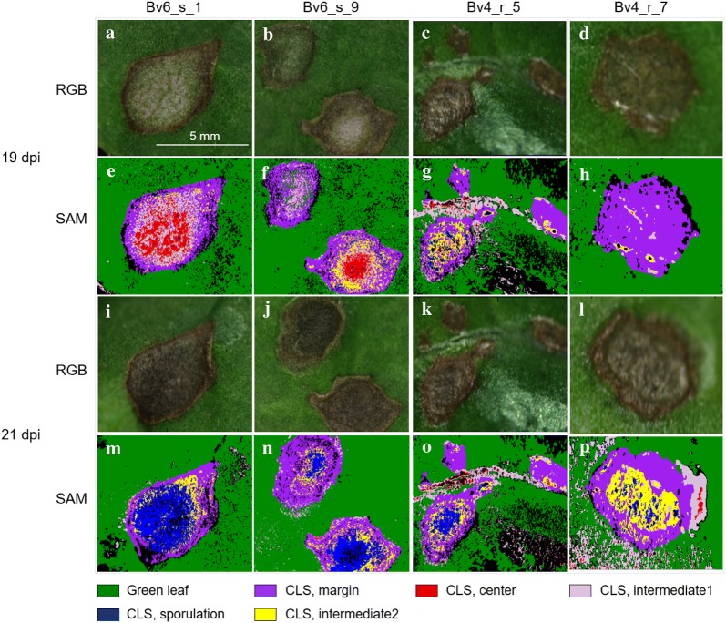
Fig. 5.
Quantification of Cercospora leaf spot (CLS) components from leaves of two sugar beet genotypes differing in resistance to CLS by supervised classification using the spectral angle mapper (SAM) algorithm. For genotypes Bv6_s (susceptible) and Bv4_r (partially resistant), two leaf areas with lesions were analyzed by recording hyperspectral images which were classified by using reference spectra (given in Fig. 4) in SAM 19 dpi (before sporulation) and 21 dpi (after induction of C. beticola sporulation); RGB representations from hyperspectral recording 19 dpi (a–d) and 21 dpi (i–l), SAM results 19 dpi (e–h) and 21 dpi (m–p). CLS intermediate 1 and CLS intermediate 2 refer to non-sporulating areas before and with sporulation, respectively

