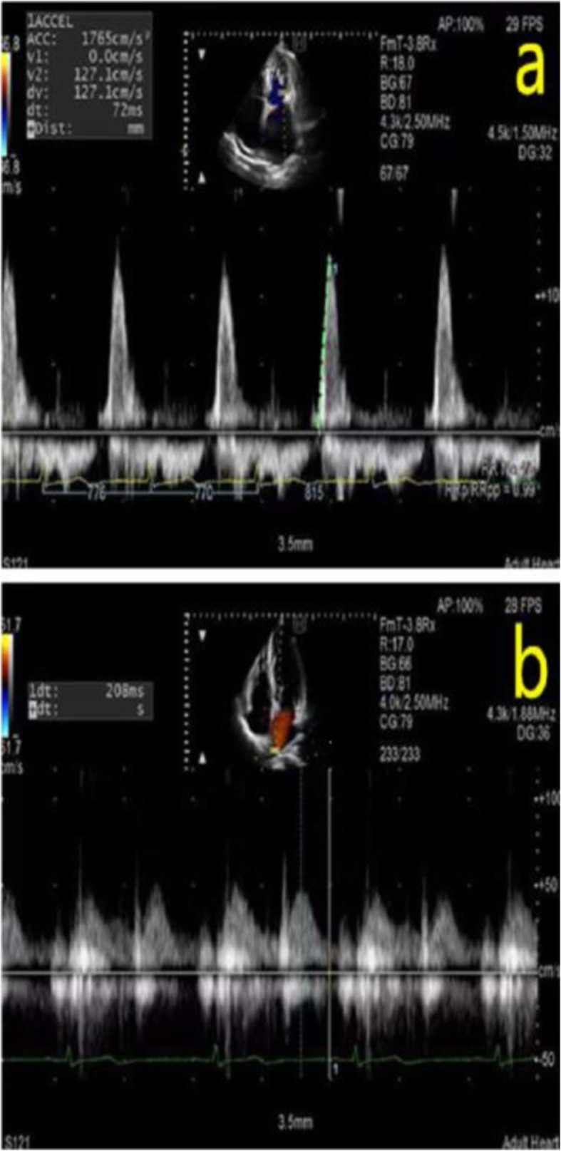
Fig. 1.

a Transmitral velocity was tracked and peak acceleration of the early transmitral flow peak velocity was measured. b. The flow of the right pulmonary vein was confirmed with the help of color doppler ultrasound in the apical four-chamber view and we measured deceleration time of pulmonary venous diastolic velocity
