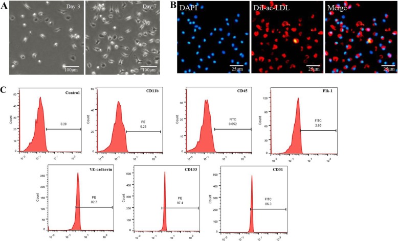
Fig. 1.
Characterization of endothelial progenitor cells (EPCs). a Morphology of EPCs was observed under a microscope. Bar, 100 μm. b DiI-ac-LDL (red) could be taken up by EPCs (the nucleus stained with DAPI, blue) as visualized through immunofluorescence staining. Bar, 25 μm. c Immunophenotypic analyses of EPC surface markers by flow cytometry

