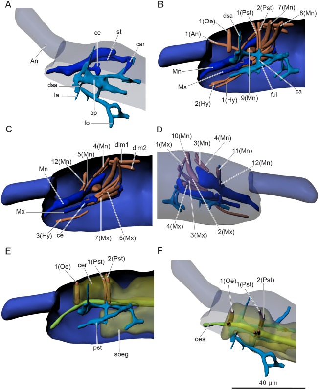
Figure 3. Anatomy of head in Mesaphorura sylvatica, 3D.
(A, F) Dorsolateral view, (B, C, E) lateral internal view, (D) lateral external view. An, antennae; bp, body of pseudotentorium; ca, connecting arm; car, cardo; ce, chitinous expansion; cer, cerebrum; dsa, dorsal suspensory arm; fo, foot; ful, fulcrum; la, lateral arm; Mn, mandible; Mx, maxillae; oes, oesophagus; pst, pseudotentorium; soeg, suboesophagal ganglion; st, stipes. Musculature see text. Paired structures (maxillae, mandible, muscles, except: 1(Oe), 1(Pst), 2(Pst) in E–F) are shown on the right side only.

