Figure 1.
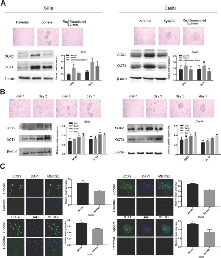
Oncosphere from cervical cancer cells exhibits high expression of stem cell markers.
Notes: Phase contrast photomicrographs and Western blot detection of the expression of SOX2 and OCT4 proteins (A) in parental adherent SiHa and CaSki cells (left), oncospheres in low-adherence culture (middle), and re-differentiated oncospheres that returned to adherent culture with 10% FBS medium (right); and (B) at days 1, 3, 5, and 7 from parental adherent SiHa and CaSki cells through development of spheroid clusters to oncospheres. (C) Immunofluorescence images of SOX2 and OCT4 in parental adherent SiHa and CaSki cells and oncospheres using an anti-SOX2/OCT4 (green) antibody and DAPI staining (blue) to indicate the cell nucleus. Images on the left show cells stained with anti-SOX2/OCT4, images in the middle show cells stained with DAPI, and images on the right are merged anti-SOX2/OCT4 and DAPI. All of the contrast of images were taken under the same conditions. *P<0.05 and **P<0.01 when comparing experimental and control groups (two-tailed Student's t test).

