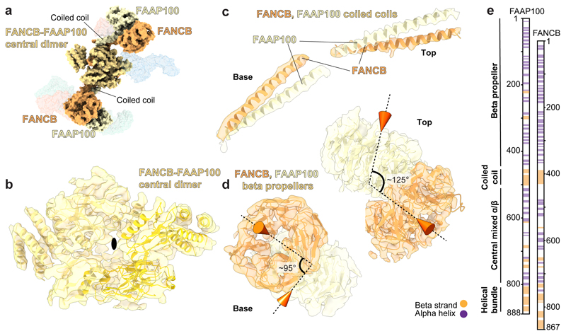
Fig. 2. The molecular scaffold of the FA core complex includes a dimer of FANCB-FAAP100 heterodimers.
a Surface representation of the FA core complex model highlighting FANCB and FAAP100. FANCB is in orange; FAAP100 is in yellow; and regions where we are unable to distinguish FANCB and FAAP100 are in yellow-orange. b-d Models of FANCB and FAAP100 subunits in cartoon representation placed into the cryoEM map. In panel (b), a black oval marks the pseudo two-fold symmetry axis. The two symmetry-related copies are shown as different shades of yellow in the model. In panel (d), cones indicate the orientations of the central pores of the beta propellers and the angles between them are given. e Proposed domain organization of FANCB and FAAP100 showing their structural similarity.

