Figure 2.
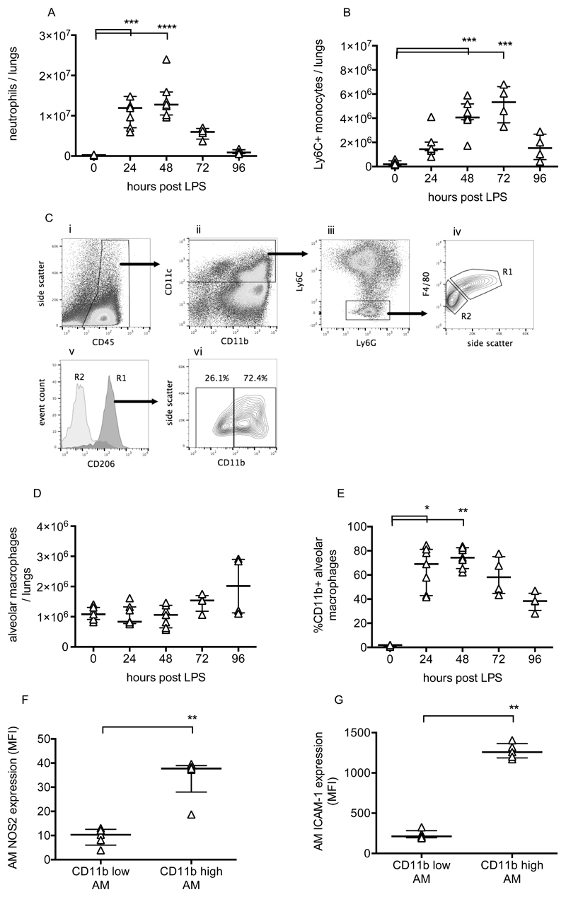
Leukocyte changes during LPS-induced lung injury. Lung tissue neutrophils (A) and Ly6C+ inflammatory monocytes (B) were quantified using flow cytometry of lung cell suspensions. Identification of resident alveolar macrophages in lungs 48 hours after LPS treatment is shown in panel C. Macrophages were identified as CD45+ (Ci), CD11c+ (Cii), Ly6C and Ly6G low (Ciii), and F4/80 high (Civ, gate R1). Gate R1 cells were also confirmed as being CD206 (mannose receptor) positive (Cv), while no other cells were. Panel Cvi demonstrates that these cells, definitively identified as resident alveolar macrophages, existed in both CD11b positive and negative populations. Panel D shows alveolar macrophage number in lung tissue, and panel E shows % of resident macrophages positive for CD11b expression. Expression of nitric oxide synthase 2 (NOS2; panel F) and ICAM-1 (Panel G) were evaluated in CD11b+ and CD11b- alveolar macrophages at 48 hours after LPS. N=4-7 for each readout at each time point, so all were treated as non-normally distributed data and are displayed as individual points with median and interquartile range. Data in Panels A, B, D & E were analysed by Kruskal-Wallis test followed by Dunn’s multiple comparisons test; *p<0.05, **p<0.01, ***p<0.001, ****p<0.0001 vs 0h (untreated mice). Data in Panels F & G were analysed by Mann-Whitney U-test; **p<0.01. Exact p-values were as follows: Panel A - 24h p=0.0008, 48h p<0.0001, 72h p=0.2191, 96h p>0.9999; Panel B - 24h p=0.1213, 48h p=0.0003, 72h p=0.0003, 96h p=0.4951; Panel D - 24h p>0.9999, 48h p>0.9999, 72h p=0.4571, 96h p=0.4571; Panel E - 24h p=0.0121, 48h p=0.0015, 72h p=0.1059, 96h p>0.9999; Panel F p=0.0079; Panel G p=0.0079.

