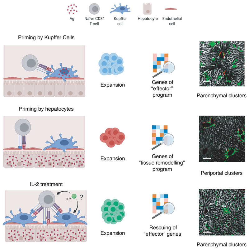
Extended Data Fig. 10. Graphical abstract summarizing the paper’s main findings.
(Top panels) Priming by Kupffer cells – that are not natural targets of HBV – leads to differentiation into bona fide effector cells that form dense, extravascular clusters of rather immotile cells scattered throughout the liver. (Middle panels) Priming by hepatocytes – the natural targets of HBV - leads to local activation and proliferation but lack of differentiation into effector cells; these dysfunctional cells express a unique set of genes including some belonging to GO categories linked to tissue remodelling and they form loose, intravascular clusters of motile cells that coalesce around portal tracts. (Bottom panels) CD8+ T cells primed by hepatocytes can be rescued by IL-2 treatment.

