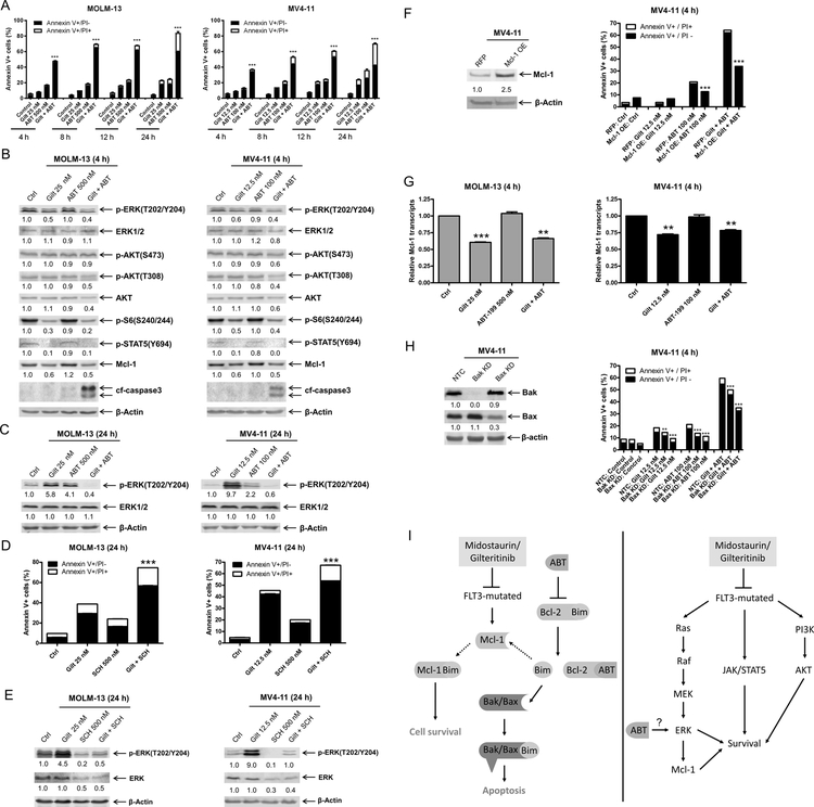
Figure 6. Gilteritinib enhances venetoclax-induced apoptosis through downregulation of Mcl-1.
(A) MOLM-13 and MV4–11 cells were treated with gilteritinib and venetoclax, alone or in combination, for up to 24 hours. Annexin V-FITC/PI staining and flow cytometry analyses results are shown. ***indicates p<0.001. (B) MOLM-13 and MV4–11 cells were treated with gilteritinib and venetoclax, alone or in combination, for 4 hours. Whole cell lysates were subjected to Western blot analyses. Representative blots are shown; densitometry measurements, shown below the corresponding blot, were normalized to β-actin and expressed as fold change compared to vehicle control. (C) MOLM-13 and MV4–11 cells were treated with gilteritinib and venetoclax, alone or in combination, for 24 hours. Representative Western blots are shown; densitometry measurements, shown below the corresponding blot, were normalized to β-actin and expressed as fold change compared to vehicle control. (D&E) MOLM-13 and MV4–11 cells were treated with gilteritinib alone or in combination with the ERK-selective inhibitor SCH772984 for 24 hours. Flow cytometry (panel D) and Western blot analyses (panel E) are shown. ***indicates p<0.001. Densitometry measurements, shown below the corresponding blot, were normalized to β-actin and expressed as fold change compared to vehicle control. (F) Lentiviral OE of RFP control and Mcl-1 were performed as described in the ‘>Methods’ section. Whole cell lysates were subjected to Western blot (upper panel). Densitometry measurements, shown below the corresponding blot, were normalized to β-actin and expressed as fold change compared to vehicle control. Overexpression cells were treated with gilteritinib and venetoclax, alone and in combination, for 4 hours. Flow cytometry analyses results are shown (lower panel). ***indicates p<0.001. (G) MOLM-13 and MV4–11 cells were treated with gilteritinib and venetoclax, alone and in combination, for 4 hours. Total RNA was isolated and Mcl-1 transcripts were determined by real-time RT-PCR by using TaqMan probes. **indicates p<0.01 and ***indicates p<0.001. (H) Lentiviral shRNA knockdown of Bak and Bax were performed as described in the ‘>Methods’ section. Whole cell lysates were subjected to Western blot (left panel). Densitometry measurements, shown below the corresponding blot, were normalized to β-actin and expressed as fold change compared to vehicle control. shRNA knockdown cells were treated with gilteritinib and venetoclax, alone and in combination, for 4 hours. Flow cytometry analyses results are shown (right panel). ***indicates p<0.001. (I) Inhibition of FLT3 synergizes with venetoclax via two proposed mechanisms in FLT3-mutated AML cells. Inhibition of FLT3 downregulates Mcl-1. Venetoclax frees Bim from Bcl-2, but also that this Bim can then be sequestered by Mcl-1, and therefore that joint Bcl-2 inhibition and Mcl-1 downregulation/inhibition is required for effective induction of apoptosis (left panel). Venetoclax potently induced p-ERK, which is abrogated by the addition of midostaurin or gilteritinib, leading to enhanced cell death.

