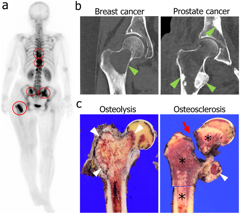
Fig. 4.
Clinical presentation of bone metastases. a A full-body bone scan in a breast cancer patient using technetium-99m shows multiple bone metastatic lesions in the spine, pelvis, and femur (red circles). b CT hip images demonstrate distinct differences in bone metastasis patterns based on the origin of cancer cells. Osteolytic remodeling is seen in a breast cancer bone metastasis (left, green arrowhead), whereas osteosclerotic remodeling is seen in prostate cancer bone metastases (right, green arrowheads). c A gross specimen shows an example of osteolytic remodeling in renal cell carcinoma with cortical erosion and loss of cancellous bone (left, white arrowheads). Osteosclerotic remodeling in prostate cancer (right, asterisks) is marked by bone production in the lesser trochanter (right, white arrowhead) and a pathologic femoral neck fracture (right, red arrow). Clinical images were approved to present in this article by the Institutional Review Board in Gunma University Hospital on October 7, 2015 (Registration no. 15-58)

