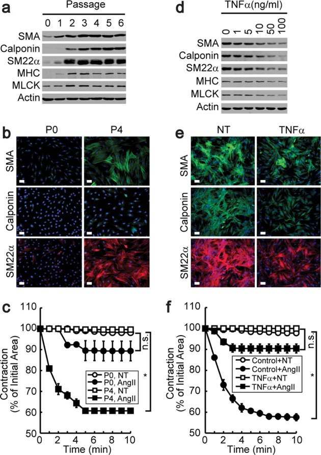
Fig. 1. Phenotypic conversion of contractile VSMCs by TNFα.

Synthetic VSMCs isolated from rat aortic tissue fragments were explanted onto laminin-coated plates. The expression of contractile marker genes was assessed by western blot analysis (a) or immunocytochemistry (b) at the indicated passages. Magnification, 40×. Bar, 100 μm. c Cells from each passage were embedded in collagen gel beads and stimulated with AngII. Time-lapse images were recorded digitally, and contractions are expressed as the percentage of the initial area (n = 3). *P < 0.05 compared with the no- treatment (NT) group. n.s., not significant. P4-stage VSMCs were stimulated with TNFα for 4 days at the indicated doses, and the expression of contractile marker genes was assessed by western blot analysis (d) and immunocytochemistry (e). Magnification, 40×. f Contractile VSMCs were stimulated with TNFα for 4 days, and AngII-induced collagen gel contraction was analyzed as described above (n = 3). *P < 0.05 compared with the no-treatment (NT) group. n.s. not significant. The results are presented as the means ± SEM. One-way ANOVA and Tukey’s multiple comparison test were used to determine the P values. The asterisks indicate statistical significance (P < 0.05).
