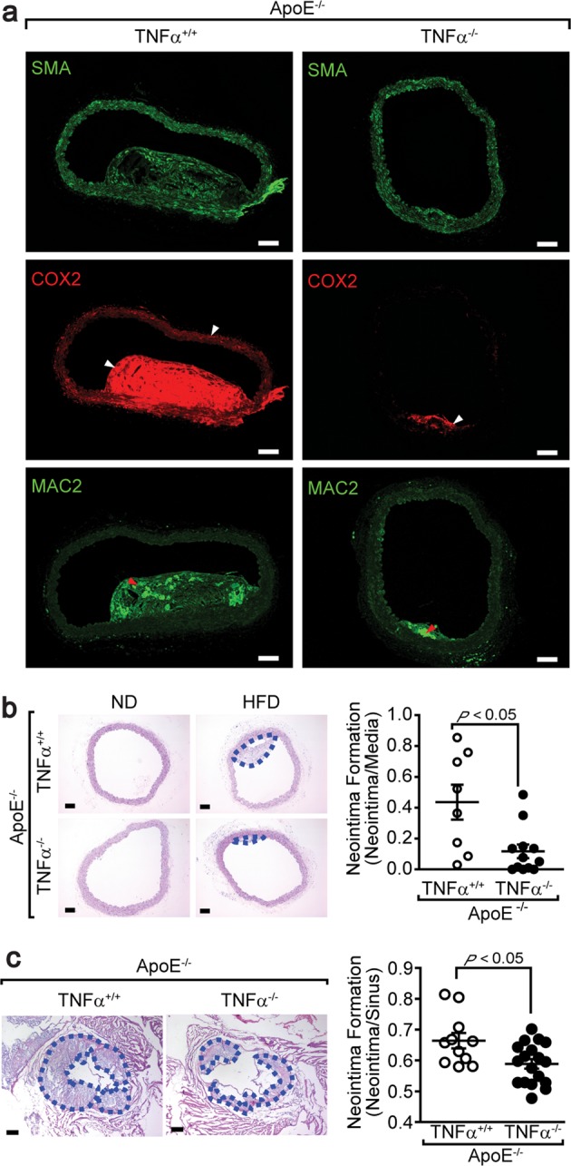
Fig. 5. Suppression of atherosclerosis in mice lacking TNFα.

a Mice lacking both ApoE and TNFα were fed a high-fat diet for 15 weeks. Aortic tissues were isolated and stained with the indicated antibodies. The white and red arrows indicate COX2 expression and macrophages, respectively. Bar, 25 μm. The mice were fed either a normal or high-fat diet for 15 weeks. Aortas (b) (ApoE−/−TNFα+/+ mice, n = 8; ApoE−/−TNFα−/− mice, n = 12) and aortic sinuses (c) (ApoE−/−TNFα+/+ mice, n = 11; ApoE–/–TNFα–/– mice, n = 18) were stained with hematoxylin and eosin, and the neointimal areas were measured. The dashed blue lines indicate neointimal tissues. Bar, 50 μm. The results are presented as the means ± SEM. Unpaired Student’s t test (two tails) was used to determine the P values.
