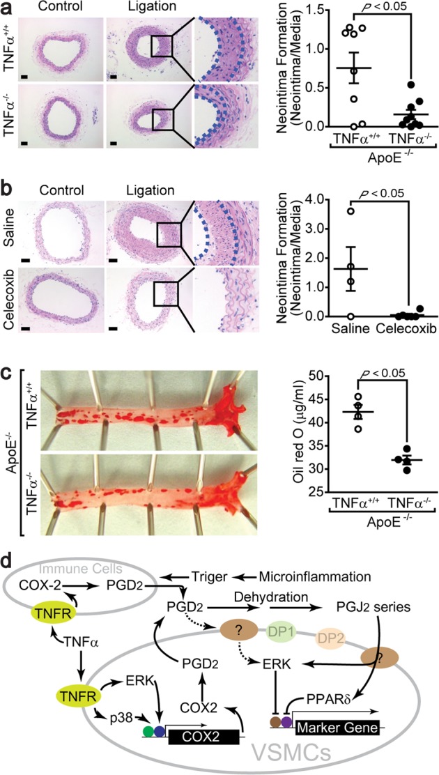
Fig. 6. Suppression of neointima formation by COX2 inhibition.

a The carotid arteries of mice lacking TNFα were ligated for 4 weeks (ApoE−/−TNFα+/+ mice, n = 8; ApoE−/−TNFα−/− mice, n = 9). Representative photomicrographs of cross sections of carotid arteries stained with hematoxylin and eosin are shown. The dashed line defines the medial and neointimal layers. Bar, 25 μm. b The mice were fed a diet containing a selective COX2 inhibitor (celecoxib) for 2 weeks before carotid artery ligation and fed a diet containing celecoxib for an additional 4 weeks (saline, n = 4; celecoxib, n = 6). Aortic tissues were isolated, and cross sections from 2 mm distal to the ligation were stained with hematoxylin and eosin. The dashed lines define the medial and neointimal layers. Bar, 25 μm. c The mice were fed a high-fat diet for 15 weeks, abdominal aortas were isolated and stained with oil red O, and lipid accumulation was quantified by extracting oil red O with isopropyl alcohol (n = 4). d A schematic representation of the PGD2-mediated phenotypic modulation of VSMCs by PPARδ. The results are presented as the means ± SEM. Unpaired Student’s t test (two tails) was used to determine the P values.
