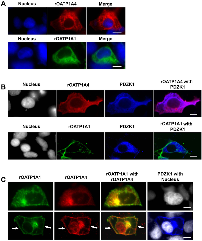
Figure 2: Subcellular localization of rOATP1A1 and rOATP1A4 when expressed singly and together in the presence or absence of PDZK1.
A. Human embryonic kidney (HEK) 293 cells were transfected with plasmid encoding sfGFP-rOATP1A1 or mRFP-rOATP1A4. Two days after transfection cells were fixed and stained with primary rOATP1A4 antibody followed by Cy5 secondary antibody and imaged by confocal microscopy with a 60x objective. Expression of sfGFP-rOATP1A1 is in green, mRFP-rOATP1A4 in red, and Hoechst 33342 stained nuclei in blue. Top: Representative images of mRFP-rOATP1A4 transfected HEK293 cells. Bottom: Representative images of sfGFP-rOATP1A1 transfected HEK293 cells. Both proteins are largely intracellular. Scale bars = 10μm. B. HEK 293 cells expressing sfGFP-rOATP1A1 or mRFP-rOATP1A4 were transfected with pFLAG-PDZK1. Cells were fixed and labeled with rOATP1A4 and PDZK1 antibodies. Expression of sfGFP-rOATP1A1 is in green, mRFP-rOATP1A4 in red, PDZK1 in blue, and Hoechst 33342 stained nuclei in gray. Images were taken by confocal microcopy with a 60x objective. Top: Representative images of mRFP-rOATP1A4 and PDZK1 transfected HEK293 cells Bottom: Representative image of sfGFP-rOATP1A1 double transfected with PDZK1 in HEK293 cells. rOATP1A1 but not rOATP1A4 was distributed on the plasma membrane in the presence of PDZK1. Scale bars = 10μm. C. HEK 293 cells were transfected with plasmid encoding sfGFP-rOATP1A1, mRFP-rOATP1A4, and/or pFLAG-PDZK1. Cells were fixed and immunofluorescence labeled with rOATP1A4 and PDZK1 antibodies. Expression of sfGFP-rOATP1A1 is in green, mRFP-rOATP1A4 in red, PDZK1 in blue and Hoechst 33342 stained nuclei in gray. Images were taken by confocal microcopy with a 60x objective. Top: Representative images of mRFP-rOATP1A4 and sfGFP-rOATP1A1 double transfection in HEK293 cells. Bottom: Representative image of triple transfection with mRFP-rOATP1A4, sfGFP-rOATP1A1 and pFLAG-PDZK1 in HEK293 cells. White arrows point to plasma localization of the proteins. Scale bars = 10μm.

