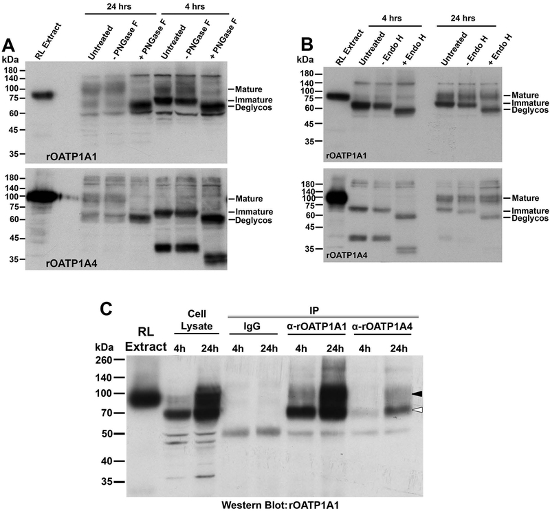
Figure 5: Assessment of the glycosylation state and interaction of rOATP1A1 and rOATP1A4 in stably transfected HeLa cells after induction of expression with zinc.
Panels A and B: HeLa cell lines expressing rOATP1A1 or rOATP1A4 under a metallothionein promoter were prepared and incubated for 4 or 24 hours, as indicated, in the presence of 150 μM zinc to initiate transporter expression. Cell lysates were prepared and were untreated or pretreated with buffer with or without PNGase F (panel A) or Endo H (panel B) prior to running immunoblots with antibody to rOATP1A1 or rOATP1A4 as indicated. A lane of rat liver homogenate that had been extracted with 0.1 M Na2CO3 (RL Extract) was included in each immunoblot to confirm antibody specificity. Positions of fully glycosylated, partially glycosylated, and fully deglycosylated OATPs are indicated on the right side of each immunoblot as mature, immature, and deglycos respectively. Panel C: Cell lysates from doubly transfected HeLa cells incubated in zinc for 4 or 24 hours were subjected to immunoprecipitation with non-immune IgG or antibody against rOATP1A1 or rOATP1A4 as indicated and Western blot was performed to detect rOATP1A1.

