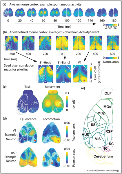
Figure 3.
Characteristics of global cortical activity in mice. (a) Spontaneous activity in the mouse cortex during quiet wakefulness recorded with wide-field VSD imaging (adapted from Ref. [34]). Observed activity is predominantly bilaterally synchronous. (b) Activity in anesthetized mouse cortex recorded with wide-field calcium imaging (adapted from Ref. [37•]), revealing globally propagating slow waves, similar to those in humans. The average of many detected ‘Global Brain Activity’ events (upper row). Individual moments in this stereotyped wave resemble (arrows) the overall seed pixel correlation maps for different seed pixel locations (lower row). (c) Activity related to movement of the mouse’s face or body dominates global dynamics, accounting for much more unique explained variance in wide-field calcium images than factors related to goal-directed behavior, over all cortical areas measured (adapted from Ref. [41•]). Colormap represents the cross-validated unique variance explained by each group of predictors (those related to the task, or those related to other movements). (d) Measurements of individual neurons with simultaneous wide-field calcium imaging allows linking local and global dynamics. Each map represents a seed neuron correlation map: the correlation between spiking activity of an individual neuron recorded electrophysiologically (at the location of the red circle) and calcium fluorescence across cortex. These data reveal that individual neurons have distinct patterns, and that these correlations can vary strongly with behavioral state, from quiescence to locomotion (adapted from Ref. [44••]). (e) For orientation, a map of the left hemisphere of the mouse brain, viewed from above. The outlines of these cortical areas are superimposed in gray over the left-most image of each preceding panel (superpositions done manually and approximately, for general guidance). Abbreviations: OLF, olfactory bulb; MOs, secondary motor cortex; MOp, primary motor cortex; SS, somatosensory areas; RSP, retrosplenial cortex; AUD, auditory areas; VIS, visual areas; SC, superior colliculus; IC, inferior colliculus. Atlas data from the Allen Institute for Brain Science.

