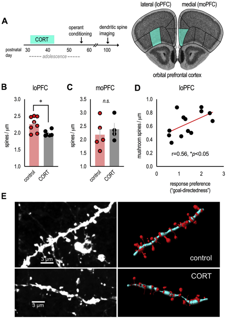
Fig. 2. Excess CORT in adolescence induces dendritic spine loss in the loPFC: Correlations with decision-making abnormalities.

(A) Experimental time (left), thy1-YFP-expressing mice from instrumental conditioning experiments in Fig. 1 were euthanized in adulthood after behavioral testing, and basilar dendrites on excitatory neurons in the loPFC (and, for comparison, moPFC) were imaged. These regions are highlighted on images from the Mouse Brain Library (Rosen et al., 2000). (B) Adolescent CORT exposure eliminated dendritic spines in the loPFC (n = 6–8/group), (C) but not moPFC (n = 4–5/group; less than loPFC due to sparseness of YFP signal in this region). (D) In the rostral loPFC, the density of mature, mushroom-shaped spines correlated with response preference ratios from Fig. 1D. n = 6–7/group. (E) Representative dendrites from the loPFC (unprocessed images at left, reconstructions at right). Bars represent means ± SEMs, symbols represent individual mice. *p ≤ 0.05. Dendritic spines were imaged and reconstructed by a single, blinded rater.
