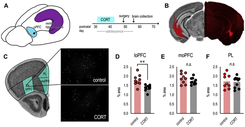
Fig. 3. Excess CORT during adolescence degrades vHC→loPFC input.

(A) Mice exposed to CORT from P31–42 received infusions of the anterograde tracer fluoro-ruby into the vHC to characterize vHC projections to the oPFC. (B) At left: The spread of fluoro-ruby in the vHC is transposed onto an image from the Mouse Brain Library (Rosen et al., 2000). At right: Representative image of fluoro-ruby in the vHC. (C) Unprocessed images of fluoro-ruby-positive axon terminal punctae in the loPFC. Comparator regions are also highlighted on an image from the Mouse Brain Library (Rosen et al., 2000). (D) Adolescent CORT exposure reduced the presence of vHC terminals in the loPFC, (E) but not the moPFC (F) or PL. n = 9–11/group. Bars represent means ± SEMs, symbols represent individual mice. **p ≤ 0.001. Punctae were imaged and enumerated by a single, blinded rater; 2 independent cohorts of mice contributed to the dataset.
