Fig. 3.
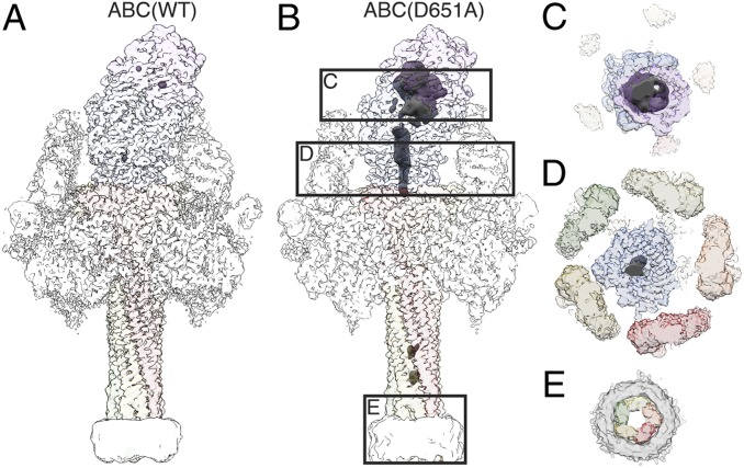
ADP-ribosyltransferase in the ABC(D651A) pore. (A and B) Reconstruction of ABC(WT) (A) and ABC(D651A) (B) with transparent surface. Density in which no atomic model was fitted, and the nanodisc were filtered to 10 Å and are shown at a lower binarization threshold. The small density blobs inside the cocoon in A and inside the TcA channel in B correspond to noise. Increasing the binarization threshold of the disordered density results in disappearance of the blobs before the density inside the cocoon disappears. See also Movie S3. (C–E) Cross-sections through ABC(D651A) at the cocoon, the TcB translocation channel, and the transmembrane region, as indicated in B. Continuous density of the ADP-ribosyltransferase is apparent in the cocoon (C) and in the narrow passage of TcB below the constriction site (D), but not in the transmembrane region embedded in the nanodisc (E).

