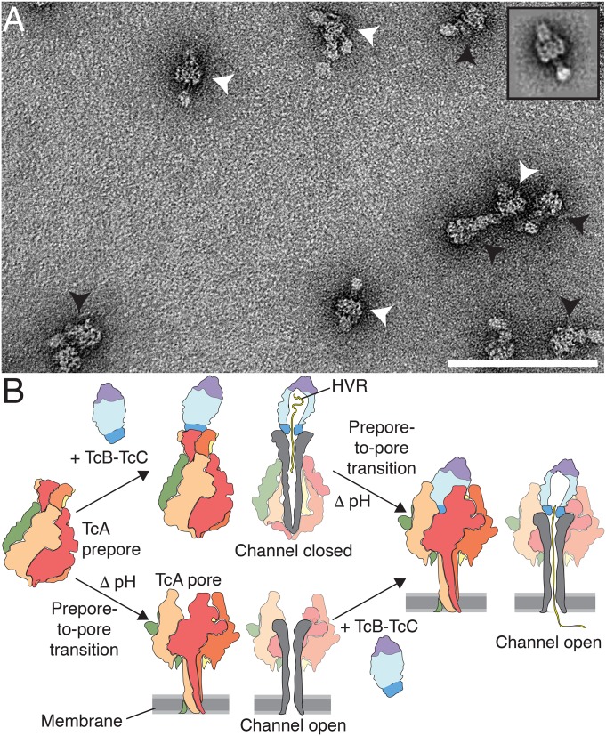
Fig. 8.
Model of 2 pathways of formation of the holotoxin pore. (A) Negative-stain electron micrograph of Tc holotoxin in nanodiscs formed by mixing TcA after the prepore-to-pore transition at pH 11 with TcB-TcC (80 and 500 nM, respectively), followed by size exclusion chromatography. Holotoxin prepores and pores are indicated with black and white arrowheads, respectively. (Inset) 2D class average of an ABC pore. (Scale bar: 100 nm.) (B) Cartoon representation of Tc holotoxin assembly, target cell interaction, and pore formation. The first pathway shows holotoxin formation with subnanomolar affinity from pentameric TcA and the TcB-TcC fusion protein (17), followed by membrane association and pH-induced prepore-to-pore transition. The second pathway shows membrane association and prepore-to-pore transition of TcA, followed by holotoxin formation of the TcA pore and TcB-TcC. Subsequently, the toxic enzyme is translocated to the target cells with its C terminus first.

