Fig. 5.
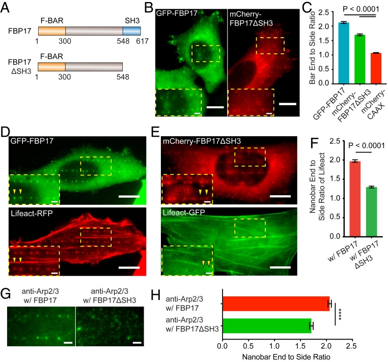
FBP17ΔSH3 reduces the curvature-dependent F-actin accumulation at nanostructures. (A) Illustrations of the full-length FBP17 and the truncated FBP17ΔSH3 plasmid constructs. (B) Both GFP-FBP17 (Left) and mCherry-FBP17ΔSH3 (Right) preferentially accumulate at the ends of nanobars. (C) Quantified nanobar end-to-side ratios of GFP-FBP17 and mCherry-FBP17ΔSH3 and mCherry-CAAX. (D) A U2OS cell cotransfected with GFP-FBP17 and Lifeact-RFP shows that both FBP17 and F-actin accumulate to the ends of nanobars. (E) A U2OS cell cotransfected with mCherry-FBP17ΔSH3 and Lifeact-GFP shows that the accumulation of FBP17ΔSH3 persists but the accumulation of F-actin at ends of nanobars is mostly eliminated. (F) Quantified nanobar end-to-side ratios of Lifeact in U2OS cells coexpressing GFP-FBP17 or mCherry-FBP17ΔSH3. (G and H) Overexpression of mCherry-FBP17ΔSH3 reduces the preference of endogenous Arp2/3 to the nanobar ends. Welch’s t test with P values indicated in C and F, ****P < 0.0001. Error bars represent SEM. (Scale bars, 10 μm [B, D, and E: full image], 2 μm [B, D, and E: zoom-in, G].)

