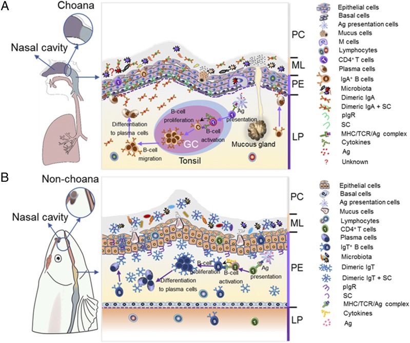
FIGURE 9.
Schematic representation of mucosal homeostasis in PM of mammals (A) and fish (B). (A) Left, In mammals, they have evolved a unique choana that connects the NC and PC, thus forming a passageway to acquire oxygen from external air. (A) Right, All mucosal regions of mammalian PC are covered by PM, which contains two main layers, an outer layer of stratified squamous epithelium (pharyngeal epithelium) and an underlying layer of dense connective tissue (LP). It is populated with both mucus-secreting cells within pharyngeal epithelium (PE) and the mucus gland within LP, which secrete mucus together into mucous layer (ML). Moreover, mammalian PM has organized lymphoid structures (i.e., tonsils) with GC, and upon pathogenic infection, it can generate activated IgA+ B cells and then migrate into the LP, where they are further differentiated into plasmablasts or plasma cells. Thereafter, IgA containing the joining J chain plasma cells is secreted by plasma cells and transported via pIgR to the ML together with mucus in control of pathogens or the recognition of microbiota. (B) Left, In contrast, because teleost fish lacks the choana, the PC is a separate compartment from the NC. (B) Right, Fish PM has two similar layers, PE and LP. Interestingly, very abundant mucus-secreting cells are instead present, which produce pharyngeal mucus directly into the ML. Moreover, IgT+ B cells are found scattered mainly in the PE, where they increase in significant numbers upon pathogenic infection. Critically, sIgT is locally generated from these IgT+ B cells, and it is transported by pIgR produced by parenchymal cells within PE into pharyngeal secretions in control of pathogens or the recognition of microbiota. Overall, in pharyngeal mucosal site, teleost sIgT and mammal sIgA (similar, but slightly different, modes of production) play a conserved role in maintaining PM homeostasis.

