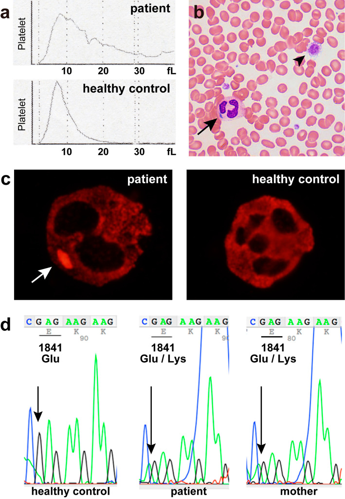
Figure 1.
(a) Platelet histograms from the patient and a healthy control. The patient’s histogram shows a broad distribution of platelets with no return to baseline, indicating the presence of abnormally sized platelets. The normal platelet histogram shows a narrow curve with a sharp peak that returns to baseline at 20 fL. (b) A peripheral blood smear shows a giant platelet (arrowhead) and Döhle-like body in the cytoplasm of a neutrophil (arrow) (Giemsa staining, original magnification ×600). (c) NMMHC-IIA localization in the neutrophils of the patient and a healthy control. NMMHC-IIA forms a cytoplasmic aggregate in a neutrophil of the patient (arrow). In the normal neutrophil, NMMHC-IIA is uniformly distributed in the cytoplasm (original magnification ×1,000). (d) A sequence analysis of the MYH9 gene reveals a heterozygous missense mutation of E1841K in the patient and his mother.

