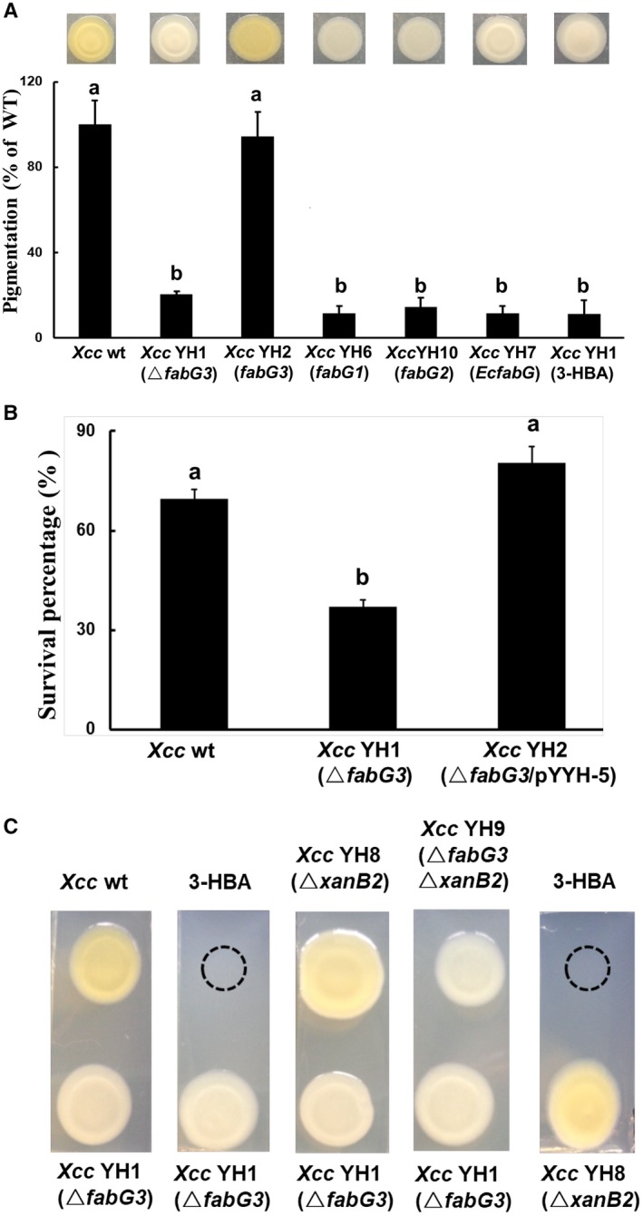
Figure 1.

The reduced xanthomonadin production and H2O2 resistance of the Xcc YH1 (ΔfabG3) strain and restoration by Xcc fabG3. (A) Quantitative analysis of xanthomonadin production in Xanthomonas campestris pv. campestris (Xcc) strains. wt, wild‐type; fabG3, complemented with plasmid pSRK‐Km harbouring the gene Xcc fabG3; Xcc fabG1, complemented with plasmid pSRK‐Km harbouring the gene Xcc fabG1; Xcc fabG2, complemented with plasmid pSRK‐Km harbouring the gene Xcc fabG2; EcfabG, complemented with plasmid pSRK‐Km harbouring the gene Escherichia coli fabG; 3‐HBA, 3‐hydroxybenzoic acid. (B) Xcc cells with an optical density of 1.0 when measured at 600 nm were collected for H2O2 treatment. After 30 min of H2O2 treatment, the CFU value of each strain was determined on NYG plates. Values shown are the means ± standard deviations from three independent experiments. Different letters indicate significant difference between treatments based on the least significant difference at P = 0.05. (C) Diffusion plate assay showing the restoration of xanthomonadin production in ΔxanB2 following exposure to Xcc YH1(ΔfabG3) or 3‐HBA, and showing the failure to restore xanthomonadin production in Xcc YH1(ΔfabG3) following exposure to wide‐type, ΔxanB2 or 3‐HBA.
