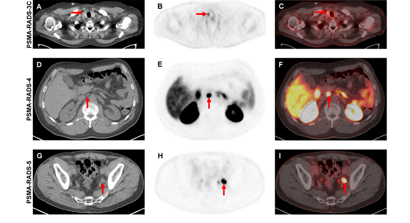
Fig. 1 –
(A–C) PSMA-RADS-3C lesion. (A) Axial attenuation-corrected CT image, (B) axial [18F]-DCFPyL PET image, and (C) axial fused [18F]-DCFPyL PET/CT image demonstrating focal radiotracer uptake in a thyroid nodule (red arrows). While this is almost certainly not a site of PCa, in the proper clinical setting this finding should be further evaluated to rule out the presence of well-differentiated thyroid cancer. (D–F) PSMA-RADS-4 lesion. (D) Axial attenuation-corrected CT image, (E) axial [18F]-DCFPyL PET image, and (F) axial fused [18F]-DCFPyL PET/CT image in a patient with recurrent PCa and a 0.6-cm lymph node (ie, not pathologically enlarged) with intense radiotracer uptake (red arrows). (G-I) PSMA-RADS-5 lesion. (G) Axial attenuation-corrected CT image, (H) axial [18F]-DCFPyL PET image, and (I) axial fused [18F]-DCFPyL PET/CT image in a patient with recurrent PCa and a 1.5-cm left pelvic lymph node (ie, pathologically enlarged) with intense radiotracer uptake (red arrows). CT = computed tomography; PET = positron emission tomography; PCa = prostate cancer; [18F]-DCFPyL = 2-(3-(1-carboxy-5-[(6-[18F]-fluoro-pyridine-3-carbonyl)-amino]-pentyl)-ureido)-pentanedioic acid.

