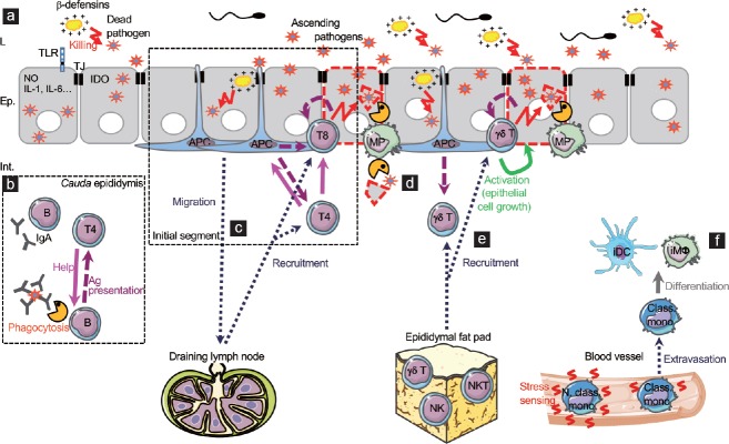
Figure 1.
Model of innate and adaptive responses to urogenital tract ascending pathogens. (a) Epididymal epithelial cells have evolved innate mechanisms that fight pathogens, of which expression of various TLRs, antimicrobial molecules (nitric oxide, IDO, β-defensins, etc.), and pro-inflammatory cytokines (IL-1, IL-6, etc.). (b) In the cauda epididymidis, interstitial B cells may be responsible for the secretion of local IgAs or may phagocytose antibody-coated bacteria and limit their dissemination in the tissue and induce specific effector T cells. Activated B cells may then classically activate helper T cells. (c) In the initial segment, the high proportion of MPs/APCs and canonical effector T cells in the tissue suggest that classical immune responses are set towards pathogens. APCs could sample circulating foreign antigens and present them to effector CD4+ and CD8+ T cells, inducing cytotoxicity of infected cells while helper T cells sustain the reaction. Depending on the population of APCs, some cells could migrate to the draining lymph node to elicit the recruitment of effector cells to the infected epididymis. (d) In case of epithelial damage due to a severe infection, some CX3CR1+CD11c+ MPs may participate in the elimination of cellular or pathogen debris. (e) The newly identified epididymal gd T cells could be activated either by APCs or directly by infected cells. Once activated, they could become cytotoxic thus participating in bacterial clearance. They are also expected to promote epithelial cell maintenance in case of cell injury following severe infections. The epididymal fat pad is suggested as a potential immune reservoir of cytotoxic cells before being recruited during infections. (f) The few monocytes identified in the epididymis are expected to sense local stress signal that induces their extravasation into the tissue and their subsequent differentiation into inflammatory MPs (iDCs and iMΦ) that temporarily sustain the immune response. Ep: epithelium; L: lumen; Int.: interstitium; TJ: tight junction; TLR: Toll-like receptor; NO: nitric oxide; IDO: indoleamine 2,3-dioxygenase; IL: interleukin; Ig: immunoglobulin; Ag: antigen; APC: antigen-presenting cell; MP: mononuclear phagocyte; NK: natural killer cell; NKT: natural killer T cell; iDC: inflammatory dendritic cell; iMf: inflammatory macrophage; Class. mono: classical monocyte; N. class. mono: nonclassical monocyte. Note: parts of the Figure 1–3 use illustrations modified from Servier Medical Art, licensed under the Creative Commons Attribution 3.0 unported license. To view a copy of this license, visit http://creativecommons.org/licenses/by/3.0/ .

