
Figure 3.
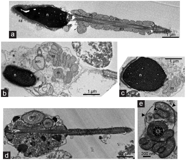
Transmission electron microscopy analysis of normal and cryptorchid spermatozoa. (a) Longitudinal section of spermatozoa from control man presents 13–14 mitochondrial gyres in a regular arrangement. Longitudinal sections of spermatozoa from cryptorchid men manifesting (b and c) partly packaged, (d) unpackaged mitochondrial gyres and (b and d) aberrant mitochondria in large cytoplasmic droplet. Note the nuclei with an uncondensed chromatin in b and c, (c) partly detached nuclear envelope (asterisk), or (b) those (arrow) forming swirling invaginations into the cytoplasm, and (d) seemingly acephalic spermatozoon. (e) Cross-section passing through the connecting piece/midpiece of spermatozoon reveals both intact and damaged mitochondria (arrowhead) gyred around the correct axoneme. a: acrosome; ra: reacted acrosome; m: mitochondria; am: aberrant mitochondria; n: nucleus; si: swirling invaginations.