Fig. 6.
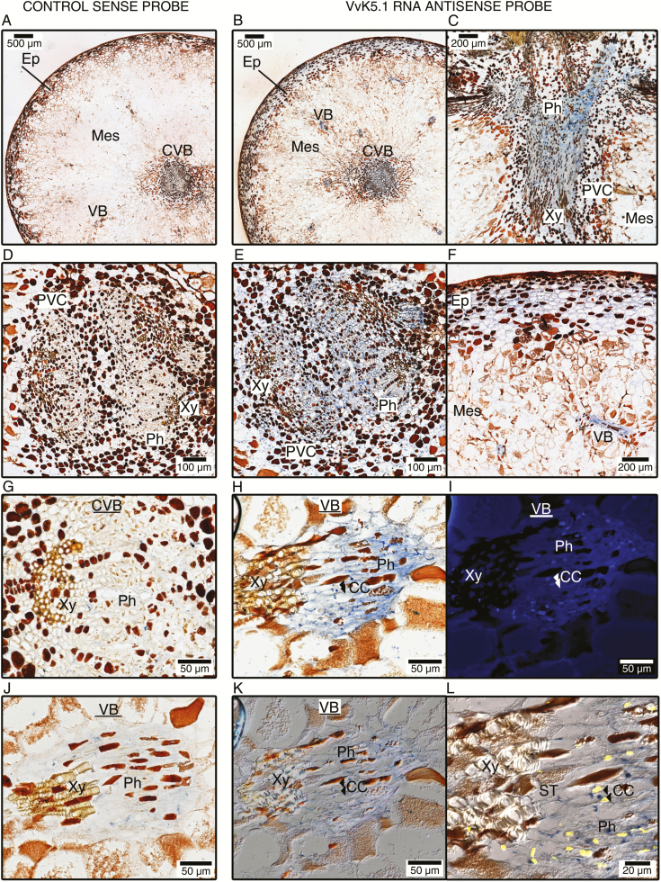
In situ localization of VvK5.1 transcripts in berries during ripening. Longitudinal and equatorial sections were hybridized with the VvK5.1 RNA sense probe as negative control (left column: A, D, G, J) or VvK5.1 RNA antisense probe (right column: B, C, E, F, H, I, K, L). Sections probed with sense probes did not show any significant signal. Sections hybridized with RNA antisense probe showed positive blue signals in ripening berries. Intense blue signals were specifically found in the phloem (B, C, E), perivascular cells (B, C, E), and, to a lesser extent, in the epicarp cells (F). DAPI staining was performed after in situ hybridization of a longitudinal section revealed a vascular bundle (H), in order to identify companion cells via their nucleus and to distinguish them from enucleated phloem sieve tubes. The section stained with DAPI was observed by fluorescence microscopy to localize cell nuclei (I), and by DIC microscopy to visualize the cell walls (K). A zoomed composite picture (L) merging (H), (I), and (K) was constructed using Image J to help localize phloem companion cells (CCs) and enucleated sieve tubes (STs). As an example, the locations of two CCs are indicated by double arrows in (H), (I), (K), and (L). Note that these two cells display a positive blue signal after in situ hybridization (H and L). CC, companion cells; CVB, central vascular bundle; Ep, epicarp; VB, vascular bundle; Mes, mesocarp; Ph, phloem; PVC, perivascular cells; ST, sieve tubes; Xy, xylem.

